In This Chapter
 Looking at the basic structure and functions of the cardiovascular system
Assessing cardiovascular findings and identifying acute coronary syndrome
Taking care of potential cardiovascular problems
More Americans experience cardiovascular-related medical conditions and emergencies than any other disease. That’s the reason why the national EMT examination makes this condition a section unto itself.
It has become clear that emergency medical services (EMS) can make a big difference in the big daddy of cardiovascular disease, the acute myocardial infarction (AMI). Early detection of an AMI, along with safe, rapid transport to a receiving facility of heart attack victims quickly reduces the likelihood of death and promotes a better life long after the event is over.
If AMI is the big daddy, then cardiac arrest is the big momma. Sudden cardiac arrest used to be almost always fatal. However, with early, high-quality CPR and rapid use of automated external defibrillators, EMTs are part of the chain of survival that can reduce the patient’s chance of dying.
There are many other causes of chest pain that this chapter explores. Many have signs and symptoms of an AMI. It’s not absolutely critical that you can tell the difference all the time; what matters most is making sure that you give each patient the benefit of the doubt as you perform your assessment. In this chapter, you get the basics on the cardiovascular system, discover normal and irregular cardiovascular findings, and figure out how to handle a number of cardiovascular problems.
Checking Out the Cardiovascular System
The cardiovascular system is broken down into three broad areas: the heart, the vasculature, and the blood. They interact closely to be able to create enough pressure in the system to produce perfusion (more commonly known as circulation).
Surveying major structures
The heart is the sophisticated pump that powers the cardiovascular system (see Figure 10-1). Its four chambers can be divided in two ways:
- Top and bottom, or the atria and ventricles, respectively
- Right and left, or pulmonary and peripheral circulation, respectively
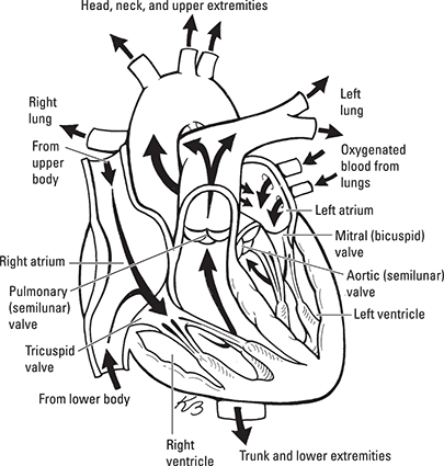
One-way valves separate the atria from the ventricles, and pulmonary circulation from peripheral circulation. This structure has the effect of forcing blood to move in one direction only, starting with blood entering the heart from the right atrium and exiting to the body through the left ventricle.
Blood is fluid that’s comprised primarily of plasma, which contains mostly water along with various salts, minerals, and proteins. Red blood cells (erythrocytes) carry most of the oxygen you need to live. White blood cells (leukocytes) fight off infection and are part of your immune system (which I describe in Chapter 11). Platelets begin the coagulation, or clotting, process when a tear in the vasculature is detected. Carbon dioxide, nutrients such as glucose, and waste such as urea are carried in the plasma.
 You’ve probably heard the phrase, “Blood is thicker than water.” Well, besides having to do with the relatives you’re born with, it is indeed true that blood has a consistency, or viscosity, that’s slightly heavier than water. The cardiovascular system relies on this viscosity to help create pressure within the vasculature.
The vasculature is the combination of pipes that the heart pumps blood into, creating pressure. The arterial side of the vasculature carries blood away from the heart, either to the lungs via the pulmonary artery to pick up oxygen or to the rest of the body via the aorta to deliver oxygen to the body in the peripheral circulation.
Arteries are made of smooth muscle and have the ability to stretch and snap back to their original shape, which helps tremendously with the flow of blood. They can also constrict and dilate, depending on the demands of the tissue for oxygen and nutrients.
Arteries divide into smaller vessels called arterioles, which divide again numerous times and finally terminate in capillary beds within the tissues. Gas exchange occurs at the capillary beds. In Chapter 9, you find out how that happens in the lungs; the process is the same everywhere else. Diffusion of CO2 and oxygen occurs based on the concentration of each gas between the capillary and the tissue cells.
The heart itself has its own vasculature. Coronary arteries branch off right where the aorta exits the left ventricle, and they supply the heart tissue, or myocardium.
Helping to return blood back to the heart is the venous system. Blood leaves the venous side of the capillary beds, collecting in venules. They, in turn, collect into veins. The veins are much more rigid than the arteries, which helps to maintain blood pressure as blood returns to the heart. Inside the veins are one-way valves that again force blood to travel in one direction.
All together, these vessels create a closed system of pipes that, with the heart acting as a pump, creates a pressure within itself (see Figure 10-2). You measure that pressure with a blood pressure cuff. By measuring the patient’s blood pressure early in your assessment, you can get a sense of how well the system is working.
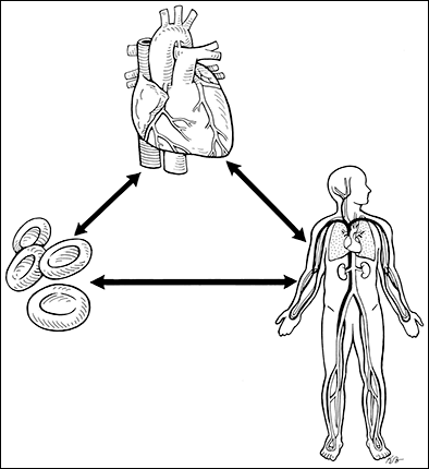
The heart, blood and vasculature make up a perfusion triangle, where all three parts interact with each other to create circulation. In reality, you measure perfusion by assessing the patient’s blood pressure.
Understanding blood pressure
One way of talking about blood pressure is with this formula:
Blood pressure (BP) = Cardiac output (CO) × systemic vascular resistance (SVR) (the size of the vasculature)
Before you panic, rest assured — you won’t be calculating BP this way! However, the formula quickly illustrates what really counts in maintaining blood pressure.
Cardiac output is the amount of blood that is sent out of the left ventricle in 1 minute. It, too, has a formula:
Cardiac output (CO) = Heart rate (HR) × stroke volume (SV) (the amount of blood squeezed out per contraction)
Again, don’t panic — no math is needed here either. But put both formulas together and this is what you get:
BP = (HR × SV) × SVR
What does all this mean? The human body controls its blood pressure through essentially one of three ways: by adjusting heart rate, stroke volume, or systemic vascular resistance. In fact, blood pressure is usually controlled via a combination of the three. Here’s an example: A patient is having an AMI that’s targeting the heart’s electrical system (see the later section “Distinguishing acute coronary syndrome from everything else” for details). The result is that the heart rate slows down. If everything were to stay the same, blood pressure would drop. But it doesn’t: The body constricts its arterial beds in the skin and other parts of the body so that SVR increases. The result is that the patient turns pale, the skin cools, and blood pressure remains near normal.
So, if the primary function of the cardiovascular system is to maintain perfusion throughout the body, it makes sense that keeping a handle on moment-to-moment changes in blood pressure controls the system. Baroreceptors in the carotid arteries do exactly that. As the receptors sense a drop in BP, they send signals to the brain that, in turn, sends signals to the body to do such things as increase heart rate, increase the strength of ventricular contractions, constrict appropriate arteries, and even decrease the amount of water being filtered by the kidneys so that more fluid stays within the bloodstream. The process is really more complicated, but in a nutshell, that’s how the body keeps its perfusion within a very narrow band of pressure.
Knowing the Cardiovascular Issues to Look for When You Assess Patients
So what are the cardiovascular system findings you should look for when assessing the patient? The following sections start with the normal findings, move to irregular findings, and wrap up with acute coronary syndrome.
Recognizing normal cardiovascular findings
You can use yourself as a “normal” picture when it comes to cardiovascular findings. Your normal resting pulse should be strong, regular, and pumping at about 60–100 beats per minute while at rest. You should be able to easily find this pulse by checking your radial artery on the thumb side of your wrist.
While you’re feeling the pulse, get a sense of how the skin looks and feels. Unless something is happening to you that I can’t see right now, I’d guess that your skin has a normal color; has a normal, warm temperature; and is dry. That’s because your skin, an organ of the body, is being adequately perfused. Now, skin can be cooler to the touch when it’s cool outside or a little sweaty when you’ve just finished a workout (or you’re panicking about your national exam), but it should return to its normal look and feel within a matter of minutes.
Your blood pressure should be roughly around 120 mm Hg or less systolic and about 80 mm Hg or less diastolic when measured at the distal bicep region of the arm. Many people have blood pressures that are higher than normal (hypertension) but live with that pressure. On the other hand, athletes and other fit folks often live with much lower BP. In general, having a lower-than-normal BP is much better than having high BP.
 Tightly connected to the cardiovascular system is the respiratory system; what happens in one is often reflected in the other. Under normal conditions, the patient’s respiratory rate should be about 12 to 20 breaths per minute without any major effort. When things start to go wrong with the cardiac system, the respiratory system tries to compensate for the issue. Flip to Chapter 9 for more information about the respiratory system.
 A 73-year-old female complains of chest tightness and difficulty catching her breath. Her blood pressure is 140/90 mm Hg, her pulse rate is 90, and her respiratory rate is 18 breaths per minute. Her skin is pale, warm, and dry; and her oxygen saturation level is 95 percent. Which of the following actions is appropriate?
(A) Lay her supine with her legs slightly elevated.
(B) Place her in a position of comfort.
(C) Administer high-flow oxygen with a nonrebreather mask.
(D) Ventilate her with a bag-valve mask.
The correct answer is Choice (B). While she may be experiencing a medical condition, her vital signs appear to be within normal limits. Keeping her comfortable will help reduce her anxiety and lower body stress. The modified Trendelenberg position, represented by Choice (A), isn’t necessary, as her blood pressure isn’t low. Her oxygen saturation level is normal, and she appears to be breathing normally; oxygen administration, Choice (C), is not indicated. She is breathing well on her own, so manual ventilations, Choice (D), aren’t necessary.
Noting when cardiovascular findings aren’t normal
Problems can arise within the cardiovascular system itself, and problems found elsewhere in the body can cause the cardiovascular system to compensate for the issue, sometimes responding so severely that it injures itself. (You find a list of common issues later in this chapter.)
When the body senses that perfusion is or may be compromised, several things happen. Heart rate increases, and the skin turns cool, pale, and clammy. Breathing speeds up too, trying to add more oxygen to the bloodstream and take out excess carbon dioxide. Combine all of these changes and the blood pressure remains as close to normal as possible, even higher than normal sometimes.
If the problem isn’t repaired, at some point the cardiovascular system can no longer compensate and begins to fail. Blood pressure falls. The heart slows. The brain, suffering from worsening hypoxia, loses its ability to maintain an alert state; the patient moves from being anxious to being confused and finds it harder to stay awake. As the condition worsens further, the patient becomes unconscious and unresponsive to a painful stimulus.
A 73-year-old female complains of chest tightness and difficulty catching her breath. Her blood pressure is 82/60 mm Hg, her pulse rate is 50, and her respiratory rate is 24 breaths per minute. Her skin is pale, cool, and dry, and her oxygen saturation level is 90 percent. Which of the following actions is appropriate?
(A) Have her sit in a chair.
(B) Administer the patient’s prescribed nitroglycerin, if available.
(C) Administer high-flow oxygen with a nonrebreather mask.
(D) Ventilate her with a bag-valve mask.
The correct answer is Choice (C). This patient’s vital signs are well outside the ordinary range. She is having difficulty breathing, and her oxygen saturation level is low, making Choice (C) necessary. Her blood pressure isn’t high enough to support sitting in a chair, Choice (A), or the administration of nitroglycerin, Choice (B). She is still breathing adequately, so manual ventilation, Choice (D), isn’t indicated.
Distinguishing acute coronary syndrome from everything else
The heart is especially sensitive to changes in perfusion. Because of its importance, the heart has a well-developed system of coronary arteries that feed the myocardial muscle. These arteries are fairly small and become easily blocked with a rupture of plaque (a layer of fat and minerals that embeds within the inner layers of an artery) or emboli (small particles of plaque) that float in from other parts of the body and lodge within the coronary artery itself. If either of these happens, the patient may experience a partial or complete loss of blood flow distal to the blockage. This occurrence marks the beginning of an acute myocardial infarction (AMI), or death of cardiac tissue.
As the cardiac tissue becomes ischemic (starved of oxygen and becoming more acidic), it sends signals back to the brain. In turn, the brain interprets these signals as the sensation of pressure, burning, tightness, sharpness, or aching. The patient may also feel nauseous, faint, lightheaded, or dizzy. Additionally, because of the close relationship of the lungs and heart, the patient can experience shortness of breath. Because of the way the nerves make their way through the body, the brain may sense related or radiating pain or discomfort to the arms, jaw, or other places in the body.
Sometimes coronary blood flow isn’t blocked by plaque or emboli. The patient may have atherosclerosis, or narrowing of the arteries that decreases blood flow to the myocardial tissue. Sometimes the arteries themselves can experience a spasm. In these situations the condition known as angina may occur. These patients have a lot of the same signs and symptoms as those with myocardial infarction. Usually someone with angina can relieve the symptoms by resting or self-administering nitroglycerin, which dilates the arteries and increases blood flow.
Angina and AMI are part of a spectrum of conditions known as acute coronary syndrome, or ACS. Recognizing these signs as early as possible is critical. Because determining the difference between angina and AMI often isn’t possible, your safest bet is to assume the worst and treat the patient quickly. Part of that treatment is to transport as quickly and safely as possible to the nearest hospital that’s capable of rapidly restoring coronary blood flow, either through angioplasty or fibrinolytic therapy.
A 50-year-old male has a sudden onset of epigastric pressure that comes on without warning. He also has pain in his jaw and feels nauseous. He has a history of ulcers and takes medication for them. He refuses your care, as he believes the discomfort is related to his ulcer history. Which of the following actions is most appropriate?
(A) Assist the patient in taking his ulcer medication.
(B) Advise the patient that he may be having a heart attack.
(C) Stay with the patient until the discomfort resolves.
(D) Begin moving the patient to your ambulance, as time is of the essence.
The answer you’re looking for is Choice (B). Although this episode may in fact be a gastrointestinal event, the symptoms also point to possible acute coronary syndrome. As an EMT, you’re not authorized to help someone with his ulcer medications, Choice (A). Staying with the patient, Choice (C), isn’t feasible, and you can’t extricate the patient, Choice (D), without his consent.
Acting on Potential Cardiovascular Problems
Like the respiratory system (covered in Chapter 9), the cardiovascular system can be affected both directly and by conditions outside of it. In the following sections, you discover problems with the heart, vasculature, and blood, and you find out how to handle a cardiac arrest.
Pump problems
In order to work effectively, the heart has to have good blood flow to its muscle via its coronary arteries and an intact electrical system that controls the rate, strength, and timing of the contractions between the atria and ventricles. Table 10-1 outlines the most common heart conditions.
Table 10-1 Conditions Affecting the Heart
|
Problem |
Signs and Symptoms |
Action Steps |
|
Acute coronary syndrome |
Described earlier in this chapter; beware of women, diabetics, and older patients having unusual signs such as shortness of breath, sudden weakness, or syncope, or no signs at all other than unexplained diaphoresis. |
Find position of comfort; evaluate need for oxygen and administer supplemental O2 as needed to maintain saturation. If patient has prescribed nitroglycerin and blood pressure is above 100 mm Hg systolic, assist with medication. If authorized, administer aspirin. |
|
Rhythm disturbances: Too fast, too slow, or too irregular |
Rates that are too fast or too slow can cause blood pressure to decrease. Radial pulses become difficult to find. Patient may experience chest pain, shortness of breath, or nausea. You may find a pacemaker or an implanted defibrillator under the skin, either in the chest or abdominal area. |
Find position of comfort. Provide oxygen as needed to maintain adequate saturation. Keep an automated external defibrillator (AED) nearby and ready for use if patient loses consciousness and pulse. Note any signs of a pacemaker or implanted defibrillator. If patient becomes pulseless, place AED pads at least 1 inch away from pacemaker or implanted defibrillator. |
|
Lethal rhythm disturbances |
Ventricular fibrillation or ventricular tachycardia that doesn’t generate a pulse. Patient is unconscious, apneic, or has gasping respirations; skin is cyanotic, cool, or diaphoretic. |
High-quality CPR with at least a 2-inch compression depth and a rate of at least 100 beats per minute. If witnessed, apply AED and follow prompts. Ventilate at a ratio of 2 breaths per 30 compressions. |
|
Cardiogenic shock |
Poor cardiac output due to AMI or other condition. Hypotension; cool, pale, and diaphoretic skin; may have chest pain associated with poor perfusion. |
Lay patient supine if possible; treat for shock by maintaining body temperature and oxygen saturation. |
|
Congestive heart failure (CHF) |
Fluid from capillary beds leaking into the alveoli in the lungs due to too-high or too-low pressure. May auscultate crackles or wheezes in lung fields. May have pedal edema from long-term CHF. If pressure is too high, may have jugular venous distension (JVD). May complain of chest pain or shortness of breath. |
If blood pressure is high, place patient in full sitting position with feet dangling if possible. Apply oxygen to maintain adequate saturation levels. Ventilate if patient is breathing inadequately. If patient is in severe respiratory distress and conscious, apply continuous positive airway pressure (CPAP). |
It is a warm, humid morning. A 78-year-old woman is attending services at her church when she faints. Church members help her to the floor. She is awake and confused as to the day and time. Her skin is cool, pale, and diaphoretic. She has a blood pressure of 82/50 mm Hg, a heart rate of 100, and a respiratory rate of 20 breaths per minute. She doesn’t have any complaints of pain or discomfort and wants to sit up. Which of the following actions is most appropriate?
(A) Assist her to a chair and perform a secondary assessment.
(B) Provide cool compresses to reduce her body temperature.
(C) Provide spinal precautions and transport to an emergency department.
(D) Place her supine on the gurney and transport to an emergency department.
The correct answer is Choice (D). Syncope, or fainting, may be caused by a variety of medical conditions. However, in this case the most serious possibility is an acute myocardial infarction, making Choice (D) the best option. The information doesn’t suggest that she is experiencing heat stroke, as Choice (B) implies, nor is there any indication that she may have injured her spine to warrant Choice (C). With her blood pressure low, sitting, Choice (A), may cause her to faint again.
Pipe problems
The body’s vasculature can develop leaks that cause fluid to leave the system quickly. On occasion, part of the vasculature can weaken, potentially causing massive failure. Table 10-2 notes common vasculature conditions.
Table 10-2 Conditions Affecting the Vasculature
|
Problem |
Signs and Symptoms |
Action Steps |
|
Aortic aneurysm/dissection |
A weakening in the aorta’s wall causing it to bulge out (aneurysm) or tear (dissection) resulting in loss of pressure and bleeding. Patient may complain of sudden tearing or knifelike pain in chest or centered between shoulder blades; may have unequal pulses in arms or legs; may experience a rapid drop in blood pressure, tachycardia, or tachypnea. In rare cases, may have a palpable abdominal mass. |
Move patient quickly but carefully; lay supine if possible. Administer oxygen to maintain saturation. Maintain body temperature. |
|
Hypertensive emergency |
Sudden rise in blood pressure over minutes to a few hours. Patient may have a rapid onset of headache; a spontaneous nosebleed; sudden ringing in the ears (tinnitus); a strong, bounding pulse; or very high blood pressure. |
Place patient in position of comfort; try to keep head elevated. Maintain oxygen saturation levels with supplemental oxygen. Attempt to control any nosebleed by pinching near base of nose and having patient lean forward. |
|
Severe infection (sepsis) |
Toxins from bacteria cause vasculature to leak fluids to surrounding tissue (third spacing). Patient may have signs of infection (fever, nausea, vomiting, diarrhea); decreasing oral fluid intake; hypotension, tachycardia, or tachypnea. Skin may be hot, dry, and pale with dark purple-colored areas where microbleeding is occurring (purpura), especially in dependent body areas of the back, buttocks, and legs. |
Place patient in supine position and monitor closely for signs of difficulty breathing. If patient is in shock, administer supplemental oxygen to maintain saturation and prevent body temperature loss. |
|
Anaphylaxis |
Massive immune response causes vasculature to dilate and leak fluid. Patient may identify source of allergen (a bee sting or peanuts, for example); may aucultate stridor or wheezing; may have hives or swelling of upper airway, hypotension, tachycardia, or tachypnea. |
Remove allergen if possible; assist patient with prescribed epinephrine autoinjector; if patient is in shock, administer supplemental oxygen to maintain saturation and prevent body temperature loss. |
A 53-year-old male is lifting several boxes at work when he feels sudden, knife-like pain in the middle of his back. He is awake and anxious, with cool, pale, and diaphoretic skin. He has a history of back surgery and hypertension. His radial pulse is weak, fast, and thready; you cannot detect pedal pulses. Of the following suspected conditions, which is most likely?
(A) Aortic abdominal aneurysm
(B) Ruptured vertebral disk
(C) Unsuspected angina
(D) Diaphragmatic hernia
The correct answer is Choice (A). The faint, fast, radial pulse and loss of pedal pulses point to a loss of blood pressure, which isn’t likely to result from a vertebral disk rupture, angina, or a diaphragmatic hernia, Choices (B), (C), and (D), respectively.
Fluid problems
If there isn’t enough blood inside the vasculature for the heart to pump, perfusion is affected. Table 10-3 notes common conditions related to blood volume.
Table 10-3 Conditions Related to Blood Volume
|
Problem |
Signs and Symptoms |
Action Steps |
|
Dehydration |
Loss of water from the blood; causes include excessive physical activity, decrease in oral intake, diarrhea, vomiting, long periods of fever (sweating), and hot environmental temperatures. Patient may complain of weakness or dizziness that may lead to a syncopal episode; cool, pale skin may be dry or wet; tachycardia or tachypnea may be present; may be hypotensive in later stages. |
If patient is in shock, administer supplemental oxygen to maintain saturation and prevent body temperature loss. Cool patient if experiencing heat stroke by removing extra clothes and fanning. In most situations, restrict oral intake. |
|
Bleeding |
Bleeding in lower GI tract may produce dark, tarry stools (melena). Patients with upper GI bleeding may have bright red or dark, coffee-ground emesis (hematemesis). If significant, patients may appear in shock with cool and clammy skin, tachycardia, tachypnea, or hypotension. |
If patient is in shock, administer supplemental oxygen to maintain saturation and prevent body temperature loss. Suction and maintain airway if patient is vomiting. |
|
Third spacing volume loss |
Fluids leak into the interstitial tissue surrounding the capillaries, as in sepsis or anaphylaxis. Patient may have signs of infection (fever, nausea, vomiting, diarrhea); decreasing oral fluid intake; hypotension, tachycardia, and tachypnea. Skin may be hot, dry, and pale with dark purple-colored areas where microbleeding is occurring (purpura), especially in dependent body areas of the back, buttocks, and legs. Massive immune response causes vasculature to dilate and leak fluid. Patient may identify source of allergen (a bee sting or peanuts, for example); may have hives, swelling of upper airway, hypotension, tachycardia, or tachypnea. |
Place patient in supine position and monitor closely for signs of difficulty breathing. If patient is in shock, administer supplemental oxygen to maintain saturation and prevent body temperature loss. Remove allergen if possible; assist patient with prescribed epinephrine autoinjector. |
A 40-year-old female is feeling faint and has difficulty breathing. You auscultate wheezing in both lung fields. Her blood pressure is 85/70 mm Hg, she has a heart rate of 110, and she is breathing 24 times per minute. Her oxygen saturation level is 85 percent. She has a prescribed inhaler for asthma and an epinephrine autoinjector for anaphylaxis. What should you do next?
(A) Assist the patient with her inhaler.
(B) Assist the patient with her epinephrine autoinjector.
(C) Complete a physical examination.
(D) Administer oxygen at 2 LPM with a nasal cannula.
The correct answer is Choice (B). Your findings are consistent with anaphylaxis, and epinephrine is the appropriate intervention for this case. Asthma, which Choice (A) points to, is unlikely to cause low blood pressure. Her oxygen level is very low, so high-flow oxygen would be a better answer than Choice (D). Although you may be able to conduct a full physical examination, Choice (C), that would come after the administration of epinephrine.
Managing a cardiac arrest
A heart that beats so weakly that it doesn’t create a pulse or doesn’t contract at all causes the condition known as cardiac arrest. Because there is no blood flow, skin becomes cold and cyanotic (blue), and the patient becomes unresponsive to all stimuli. If cardiac arrest continues for more than a few minutes, enough brain cells die to cause permanent death.
Research in the past decade has shown that effective chest compressions are the foundation of successful resuscitation. In other words, during a “working code” everything that is done revolves around the nonstop, high-quality chest compressions. Keep these points in mind:
- After checking to see whether the patient is unconscious, spend no more than 10 seconds to confirm there is no carotid pulse and breathing is absent or inadequate (gasping).
- Begin CPR with compressions, not ventilations. Immediately begin pushing on the chest, while others are assembling other equipment and preparing to ventilate.
- For adults, administer compressions at a rate of at least 100 per minute, with at least 2 inches of depth, and a full recoil of the chest during release. For pediatric patients, compress the chest at least one-third to one-half the depth of the chest.
- For adults, space ventilations so two breaths are provided after every 30 compressions. Deliver just enough to make the chest visibly rise. For two-person CPR on a pediatric patient, space ventilations so two breaths are provided after every 15 compressions.
- Rescuers should switch roles every 2 minutes or 5 cycles of compressions and ventilations, to keep compressions accurate and effective.
- Apply AED pads as soon as possible.
- As soon as the pads are applied, everyone stops and the AED is activated. Follow the prompts and make sure everyone stands clear of the patient. After the AED has analyzed, continue compressions while the AED is charging for defibrillation. When the AED is charged, stop compressions, clear the patient, and deliver the shock. Immediately afterward (or if the AED tells you that no shock is indicated), immediately begin CPR again. Do not pause to check for a pulse.
- At the end of the next 2-minute interval, look for signs of effective breathing and check for a pulse. If they are absent, immediately resume CPR. If the AED indicates a shock is needed, continue compressions while the AED charges and then clear the patient and deliver the shock.
- During two-person CPR on a child or infant, the ratio of compressions to ventilations is 15:2. This ratio allows more ventilations to be delivered to the patient.
You and three other trained crew members arrive at a park, where you see a jogger lying on the sidewalk. Two bystanders are performing CPR, with one administering chest compressions while the other is providing mouth-to-mouth ventilations. What should you do next?
(A) Stop compressions and check for a radial pulse.
(B) Stop compressions and apply the AED pads to the patient’s chest.
(C) Have other trained crew members take over compressions and ventilations at the end of a cycle.
(D) Apply AED pads during compressions and push the button to analyze when another 5 cycles of compression and ventilations are complete.
The correct choice is (C). You want to minimize interruptions to compressions, and Choice (C) accomplishes this better than Choices (A) and (B). You want to analyze the rhythm as soon as you are able, rather than waiting for 5 cycles of compressions and ventilations to be completed, as Choice (D) indicates.
Practice Questions about Cardiology and Resuscitation
The following practice questions are similar to the EMT exam’s questions about cardiology and resuscitation. Read each question carefully, and then select the answer choice that most correctly answers the question.
1. A 68-year-old male has chest pressure and shortness of breath after climbing a flight of stairs. His neighbor is worried and calls 911. The patient is alert and insists that this is an angina episode that will go away on its own. Which of the following questions would be of most benefit in verifying his assertion?
(A) Are you nauseous?
(B) Does this pain come on each time you climb these stairs?
(C) Are you prescribed nitroglycerin?
(D) Does the pain radiate to your arm or jaw?
2. A man stands up to clear the dishes after dinner. He feels a sharp, tearing sensation in the epigastric region of his abdomen, toward his back. He immediately collapses to the floor. His blood pressure is 100/84 mm Hg, his pulse rate is 108, and he is breathing 24 times per minute. His skin is pale, cool, and diaphoretic. Pedal pulses are absent. You should
(A) immobilize him to a long backboard.
(B) assist him to a sitting position.
(C) move him carefully to a gurney.
(D) administer the patient’s prescribed nitroglycerin.
3. A 72-year-old female presents with “aching” discomfort in her epigastric region that began abruptly 45 minutes ago. She reports a history of hypertension, ulcers, angina, and atherosclerosis. Her pain is a 7 on a 1–10 scale. She self-administered three of her prescribed nitroglycerin tablets, without effect. Her blood pressure is 136/90 mm Hg. You should first
(A) administer supplemental oxygen.
(B) administer another dose of the patient’s nitroglycerin.
(C) perform a full secondary assessment.
(D) determine what other medications are prescribed.
4. A 49-year-old male presents supine in bed, alert, and complaining of lightheadedness, difficulty breathing, and heavy chest pressure that radiates to his arms. His heart rate is 42, his blood pressure is 92/62 mm Hg, and he has a breathing rate of 18 times per minute. You auscultate crackles in both lung fields. You should
(A) leave the patient supine and administer supplemental oxygen.
(B) administer the patient’s prescribed nitroglycerin.
(C) sit the patient upright to assist his breathing.
(D) assist the patient’s ventilation with a bag-valve mask and oxygen.
5. A 65-year-old female is supine in bed with difficulty breathing and substernal chest pressure. She is alert and breathing 26 times per minute, with crackles auscultated bilaterally. Accessory muscle use is evident. Her blood pressure is 220/104 mm Hg, her heart rate is 90 and irregular, and she has cool, diaphoretic skin. You should
(A) assist with her breathing using a bag-valve mask and oxygen.
(B) sit her upright and administer high-flow oxygen.
(C) assist the patient with her prescribed hypertension medication.
(D) keep her supine and administer low-flow oxygen.
6. An 80-year-old male had a syncopal episode while standing during church service. Bystanders assisted him to the ground, where he presents awake, confused, and short of breath. He is pale, warm, and diaphoretic; his blood pressure is 100/70 mm Hg, and his pulse rate is 96. He is breathing 18 times per minute; his lung sounds are clear. Which of the following series of actions would be most appropriate?
(A) Sit the patient upright and assist him to a gurney, administer oxygen, and perform a full physical examination.
(B) Move the patient to a cooler spot within the church; loosen his clothing and fan him to promote cooling.
(C) Keep the patient supine and transfer him to the gurney, administer oxygen, and move toward the ambulance.
(D) Obtain a complete physical exam and past medical history, administer oxygen, and sit him upright.
7. A 74-year-old male is supine in bed. He opens his eyes when you call his name and is confused as to time and location. His skin is pale, warm, and dry. Family reports the patient has been ill, with vomiting and diarrhea for ten days, and has not been out of bed in the past 36 hours. His blood pressure is 86/64 mm Hg, he has a heart rate of 104, and he’s breathing 20 times per minute. His medical history includes diabetes, early stage lung cancer, and a myocardial infarction three years ago. Which of the following conditions is the most likely cause of his presentation?
(A) Insulin shock
(B) Septic shock
(C) Pulmonary shock
(D) Cardiogenic shock
8. A 34-year-old male presents with difficulty breathing and feeling faint. He came home from a restaurant about an hour ago, felt nauseated, and vomited several times. His pulse rate is 126, his blood pressure is 88/50 mm Hg, and he is breathing 24 times per minute. You auscultate wheezing in both lung fields. His skin is pale, cool, and moist. You should assist with the patient’s prescribed
(A) epinephrine autoinjector.
(B) multidose inhaler (MDI).
(C) nitroglycerin.
(D) insulin injection.
9. A 25-year-old female is confused and slow to respond to verbal commands. Her roommate states that the patient returned from an overseas business trip four days ago and has been experiencing nausea, vomiting, and diarrhea since. You observe yellow-colored, thin emesis in a garbage pail next to the patient’s bed. Her skin is warm, pale, and dry. She is tachycardic and tachypneic. Which of the following conditions is the most likely cause of her presentation?
(A) Gastrointestinal bleeding
(B) Dehydration
(C) Food poisoning
(D) Hypoglycemia
10. A 65-year-old male is sitting at his kitchen table feeling increasingly weak, dizzy when he stands, and nauseated over the past 6 hours. He admits to binge drinking alcohol over the past 72 hours. His skin is pale, warm, and dry. He has neither vomited nor had diarrhea, although his stool has been loose and darker than normal. His medical history includes hypertension, alcoholism, liver disease, and drug abuse. His blood pressure is 98/68 mm Hg, his heart rate is 116, and he is breathing 18 times a minute. His oxygen saturation is 92 percent. You should
(A) obtain orthostatic vitals, first when sitting and then while standing.
(B) transport the patient in a sitting position and be prepared to suction as needed.
(C) transport the patient in a left recumbent position and administer supplemental oxygen.
(D) administer high-flow oxygen and transport the patient in a sitting position.
11. You are on-scene with a 50-year-old female who is experiencing substernal chest pressure. In mid-sentence she stops talking, rolls her eyes back, and has slow, snoring respirations. Your next step is to
(A) begin deep and fast chest compressions.
(B) apply AED pads.
(C) check for a carotid pulse.
(D) ventilate twice with a bag-valve mask.
12. Bystanders are performing adequate chest compressions on an adult male found down in the middle of a food court at a shopping mall. You are with your partner and are carrying your airway equipment and an AED. You should
(A) take over chest compressions and instruct the bystanders to apply the AED pads.
(B) stop CPR and apply the AED pads yourself.
(C) have bystanders continue chest compressions while you apply the AED pads.
(D) have your partner take over compressions while you prepare the airway equipment.
13. You and your partner arrive at a public pool where lifeguards are performing CPR on a 7-year-old female who was underwater for 10 to 15 minutes. One lifeguard is performing adequate chest compressions, pausing every 30 compressions for the other lifeguard to ventilate the patient with a pocket mask. You should
(A) direct the lifeguards to continue CPR at a ratio of 15 compressions to two breaths.
(B) take over CPR and direct the lifeguards to apply the AED pads.
(C) direct the lifeguards to stop CPR and apply the AED pads.
(D) have your partner take over compressions while you apply the AED pads.
14. You are performing CPR during a cardiac arrest. You stop compressions and push the AED’s “analyze” button. After a few seconds the AED announces, “no shock indicated.” You should
(A) provide two ventilations with a bag-valve mask.
(B) begin chest compressions.
(C) check for a carotid pulse.
(D) check for breathing.
15. You finish 2 minutes of CPR on an adult patient. You detect a carotid pulse. The patient is not breathing. You should
(A) activate the AED.
(B) ventilate every 5 seconds with a bag-valve mask.
(C) begin chest compressions.
(D) place the patient in the recovery position.
16. Your patient is at her office, complaining of a headache, and “loud ringing in her ears.” She is alert, with a pulse rate of 90, blood pressure of 260/110 mm Hg, and a respiratory rate of 16 breaths per minute. She has equal strength and movement in all extremities and speaks without difficulty. She has a medical history of asthma and has an inhaler. Which of the following conditions is most likely to cause her presenting signs and symptoms?
(A) Trauma to the head
(B) Hemorrhagic stroke
(C) Ischemic stroke
(D) Hypertensive emergency
17. A 1-month-old infant is in cardiac arrest. You and your partner arrive on-scene. Which of the following actions is most appropriate?
(A) Provide chest compressions using a two-thumb, encircling hands technique, while your partner ventilates every 15 compressions.
(B) Provide chest compressions using a two-finger technique, while your partner ventilates every 30 compressions.
(C) Provide 1-inch deep chest compressions at a rate of 100 per minute.
(D) Provide ventilations with small volume over 3 seconds.
18. A 47-year-old female was asleep when she awoke with an abrupt onset of “tightness” between her shoulder blades approximately 30 minutes ago. Her condition has progressively worsened since it began; it doesn’t change with movement or body position. She hasn’t had this sensation before. She is anxious and nauseated, but doesn’t need to vomit. Her medical history includes hypertension and arteriosclerosis. Her skin is pale, warm, and dry. Her blood pressure is 136/86 mm Hg, her heart rate is 90, and she is breathing 16 times per minute. You should transport her to
(A) a trauma center with CAT scan capabilities.
(B) a hospital capable of emergency percutaneous angioplasty.
(C) her preferred hospital that contains her medical records.
(D) the closest hospital with rapid lab workup and X-ray capabilities.
Answers and Explanations
Use this answer key to score the practice questions in the preceding section. The explanations give you insight into why the correct answer is better than the other choices.
- B. A hallmark of angina is that it’s predictable, meaning the patient can tell when the pain will come on. The other questions can apply to either angina or myocardial infarction, making them less useful in verifying the patient’s assertion.
- C. This scenario has the telltale signs of an aortic dissection or rupture. He needs to be rapidly transported to a hospital where he can be evaluated for surgery. There is no indication of a trauma mechanism to indicate Choice (A), and nitroglycerin, Choice (D), may actually worsen the condition. Sitting, Choice (B), may increase the pressure on the already damaged aorta.
- A. If the first three doses of the patient’s nitroglycerin weren’t effective, it’s unlikely that any more doses, Choice (B), will help. The other choices are steps in the assessment process, but first you should apply supplemental oxygen, before moving forward, to ensure that any possible hypoxia is managed.
- A. The patient is experiencing difficulty breathing and has poor lung sounds, making oxygen necessary. The patient’s blood pressure is too low to risk having the patient sit up as Choice (C) suggests or to risk administering nitroglycerin, Choice (B). He is alert, meaning he’s able to compensate for his respiratory distress, so assisting his ventilations, Choice (D), isn’t necessary at this time.
- B. This patient is experiencing pulmonary edema secondary to congestive heart failure (CHF). Sitting her up will assist her ability to breathe. Keeping her supine, Choice (D), won’t help. While both Choices (B) and (D) include oxygen administration, the better answer is based upon the body positioning. You’re not permitted to administer antihypertensive medications, ruling out Choice (C). Her respiratory rate is adequate, so bag-valve-mask use, Choice (A), is not indicated at this time.
- C. The patient’s blood pressure is too low to have him sit up as Choices (A) and (D) suggest. Although the syncopal episode may be simply heat related, which requires cooling, Choice (B), no clear information is provided in the scenario. In addition, a myocardial infarction in elderly patients can often present as a sudden syncopal episode.
- B. The scenario suggests an infection-based condition that has steadily gotten worse. The history of lung cancer implies his immune system is compromised as well. All of this points to sepsis as the root cause of his hypotension. You would expect cool and diaphoretic skin signs for insulin shock, Choice (A), and cardiogenic shock, Choice (D). Pulmonary shock, Choice (C), is a nonsense term.
- A. The scenario suggests that the patient is experiencing anaphylaxis after eating at a restaurant. An asthma attack requiring an MDI, Choice (B), doesn’t typically cause such a dramatic change in blood pressure. On the off chance that it’s cardiogenic shock, nitroglycerin, Choice (C), is contraindicated. You aren’t permitted to inject insulin, Choice (D). Besides, that’s for cases of hyperglycemia, not hypoglycemia.
- B. The patient appears to be hypovolemic, secondary to vomiting and diarrhea. The emesis does not appear to contain blood as Choice (A) indicates; food poisoning, Choice (C), usually passes within 8–24 hours. Hypoglycemia, Choice (D), is a possibility, but it’s less likely than dehydration in this case because her skin would be cool and diaphoretic, not warm and dry.
- C. Transporting the patient on his side will help him maintain his airway; his oxygen saturation level is low, so administering oxygen will help. The patient is hypotensive; sitting the patient, as in Choices (B) and (D), may worsen his blood pressure. Because the patient is already showing signs of hypotension, orthostatic vital signs, Choice (A), won’t provide any additional information and may worsen his blood pressure as well.
- C. You need to confirm the patient is in cardiac arrest by checking the carotid pulse before beginning resuscitation, Choice (A). If she is, you can then begin chest compressions. Applying AED pads, Choice (B), will come next. At the end of 30 compressions, you’ll administer two breaths, Choice (D).
- C. If bystanders are performing good chest compressions, allow them to continue so that you can focus on applying the AED pads and activating the device. You don’t want to stop chest compressions, Choice (B), and you don’t want to tie up yourself and your partner in performing skills that are already being done correctly, Choices (A) and (D).
- A. Pediatric cardiac arrests are thought to be respiratory driven, rather than cardiac driven as in adults. In two-rescuer CPR on a child, more ventilations can be delivered using a ratio of 15 compressions to 2 breaths rather than 30 to 2. You don’t want to stop chest compressions, Choice (C), and you don’t want to tie up yourself and your partner in performing skills that are already being done, Choices (B) and (D).
- B. Chest compressions that are deep, fast, and continuous improve cardiac arrest outcomes. You want to resume compressions as soon as possible after any intervention. Choices (A), (C), and (D) all delay compressions.
- B. Your patient is in respiratory arrest. Ventilating the patient will provide much-needed oxygen during this stage. Chest compressions, Choice (C), and activating the AED, Choice (A), are not indicated at the moment. You can’t ventilate the patient while he’s on his side, contraindicating Choice (D).
- D. The scenario describes signs of a hypertensive emergency. Although it’s possible she’s having a stroke, Choices (B) and (C), her response to a Cincinnati stroke scale test (equal grips, speaks without difficulty) reduces the likelihood. You have no information to suggest a traumatic event, Choice (A), has occurred.
- A. In pediatric, two-rescuer CPR, a ratio of 15 compressions to 2 ventilations is appropriate and improves oxygenation compared to a 30:2 ratio, Choice (B). The compressions should be one-third to one-half the depth of the chest, which is at least 1½ inches deep, not 1 inch as Choice (C) indicates. Ventilation duration is similar to that of an adult, meaning that ventilations are delivered over a period of 1 second each, not 3 seconds as stated in Choice (D).
- B. This patient has all the hallmarks of a myocardial infarction, despite the absence of chest pain. In an MI, time is of the essence. The best place of care for her condition is a facility with emergency angioplasty capabilities to reopen her blocked coronary arteries.
Chapter 11
What is the most critical action EMTs can take during cardiac arrest?
Tap to revealImmediate, high-quality CPR and rapid defibrillation with an AED.
Define acute myocardial infarction (AMI).
Tap to revealA heart attack resulting from blocked blood flow to the heart muscle, causing tissue damage.
What key symptom often prompts EMS activation for cardiovascular emergencies?
Tap to revealChest pain or discomfort, sometimes accompanied by other symptoms like shortness of breath or nausea.
Practice the steps you would take if you encountered a patient with chest pain and suspected cardiac arrest.
- List the assessment steps you would perform in order.
- Identify when you would call for advanced help and begin CPR.
- Describe how you would use an AED if available.
Think of a scenario where you must decide whether a patient’s chest pain is cardiac in nature. How would you ensure you don’t miss a life-threatening emergency, and what steps would you take to provide the best care possible?
Which intervention has been shown to most significantly improve survival in sudden cardiac arrest?
Early recognition and rapid intervention during cardiovascular emergencies—especially with high-quality CPR and defibrillation—are the most effective ways EMTs can save lives.
When assessing chest pain, EMTs should always give patients the benefit of the doubt, as the signs and symptoms of serious cardiac events can overlap with other conditions.
Early detection of an AMI, along with safe, rapid transport to a receiving facility of heart attack victims, quickly reduces the likelihood of death and promotes a better life long after the event is over.
- Cardiovascular emergencies are the most common medical events for EMTs.
- Early CPR and defibrillation are central to survival in cardiac arrest.
- Chest pain should always be treated seriously and assessed thoroughly.